The Tau Code: One Test to Unmask Eight Different Brain Diseases
Eight devastating brain diseases. Eight different patterns of protein misfolding. And now, one remarkably elegant laboratory test that can tell them all apart - potentially revolutionizing how we diagnose dementia.
Listen to This Article
AI-generated discussion • ~6 min
When someone develops dementia, the cause is not always Alzheimer's disease. There is a whole family of brain diseases called tauopathies -conditions where a protein called tau misfolds and clumps together in the brain, destroying neurons. But here is the problem: different tauopathies produce different shapes of misfolded tau, and until now, the only reliable way to tell them apart was to examine brain tissue after a patient had died.
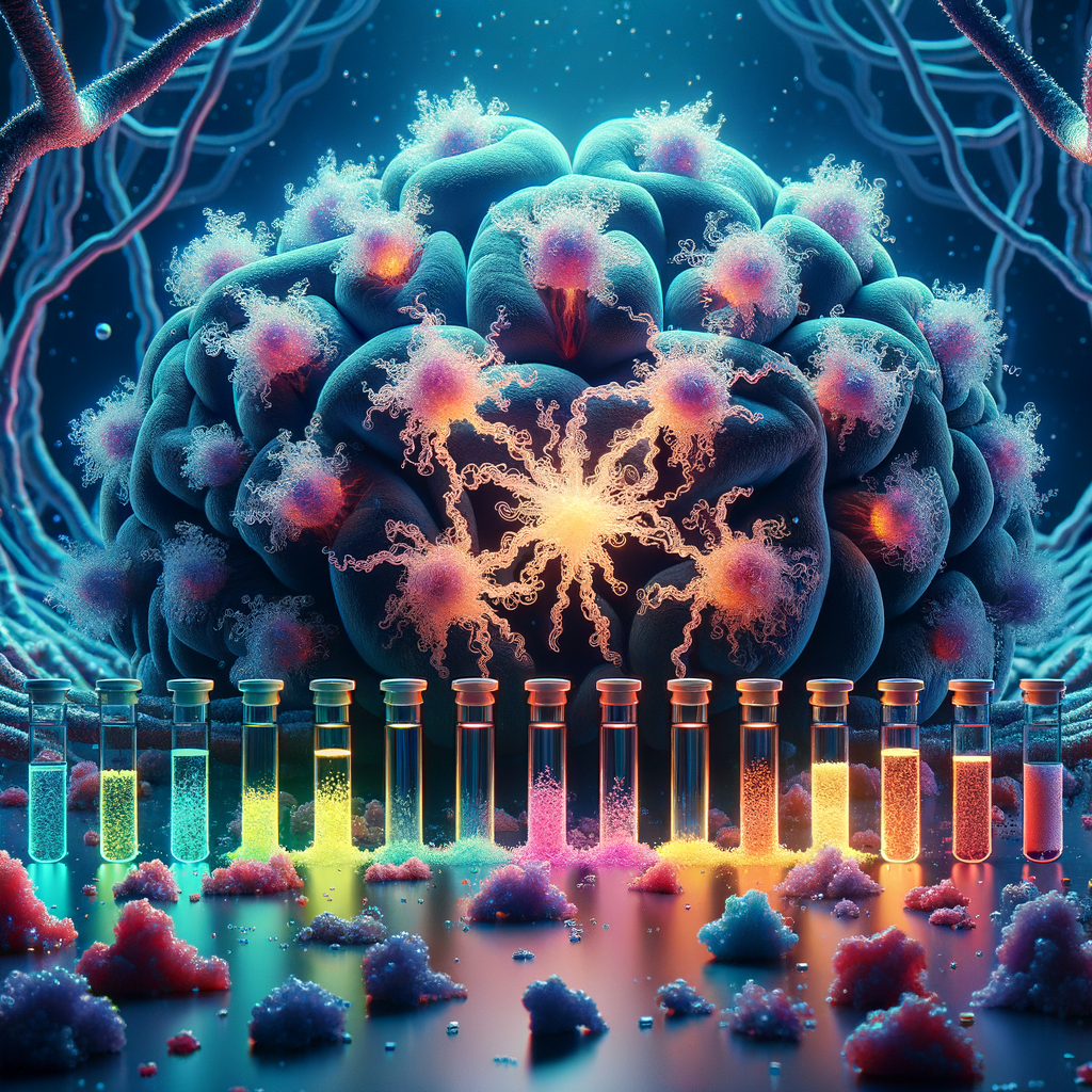
Researchers at the University of Cambridge, led by Professor Michele Vendruscolo, have developed a laboratory test that can distinguish eight different tauopathy subtypes from brain samples. The test is called RT-QuIC, and the breakthrough lies in a deceptively simple idea: by changing the salt concentration in the test, different types of misfolded tau reveal their unique signatures.
Think of it this way: imagine you have eight different types of origami cranes, all folded from the same sheet of paper but in slightly different ways. They look similar when folded up, but if you put them in water with different amounts of salt, each type unfolds and refolds at a different speed and in a different pattern. That is essentially what this test does with misfolded tau proteins. The salt acts as a molecular magnifying glass that amplifies the subtle structural differences between tauopathy strains.
The diseases the test can identify include Alzheimer's disease, Pick disease, PSP (progressive supranuclear palsy), CBD (corticobasal degeneration), AGD (argyrophilic grain disease), a genetic form called FTDP-17, and two types of GGT (globular glial tauopathy).
The key technical innovation is the use of two different tau substrates -called K12 and K11 -combined with varying sodium chloride concentrations. Some tauopathies amplify strongly with the K12 substrate, others respond better to K11, and the salt concentration determines how aggressively the amplification proceeds. By running samples through multiple conditions, the researchers built a diagnostic fingerprint for each disease.
Crucially, this test does not require heparin, which older tau amplification methods relied on. Heparin is a blood thinner that was used to kickstart tau aggregation in previous tests, but it introduced artifacts -essentially contaminating the results by forcing tau into shapes it would not naturally adopt. By removing heparin and using salt modulation instead, the Cambridge team gets a much cleaner, more faithful picture of how the disease proteins actually behave.
The research team validated their approach using brain samples from patients with confirmed diagnoses -confirmed by the gold standard of post-mortem neuropathological examination. They showed that the test reliably classified each sample into the correct disease category, with distinct amplification kinetics and fluorescence signatures for each tauopathy.
The ultimate goal is to adapt this test for living patients, using cerebrospinal fluid or even blood samples. If successful, this would transform dementia diagnosis from a post-mortem exercise into a clinical tool that could guide treatment decisions while patients are still alive. As precision medicine and targeted therapies for different tauopathies continue to develop, knowing exactly which disease a patient has becomes critical to giving them the right treatment.
Real-World Impact
Quick Takeaways
- First laboratory test capable of distinguishing eight different tauopathy subtypes from brain homogenates using a single assay platform
- Eliminates the need for heparin in tau amplification, producing cleaner and more disease-faithful results
- Uses salt concentration as a simple but powerful variable to reveal structural differences between misfolded tau strains
- Paves the way for clinical diagnostic tests using cerebrospinal fluid or blood, potentially enabling accurate dementia subtyping in living patients
The ability to accurately classify tauopathies has immediate implications for clinical trials. Many experimental therapies are designed to target specific forms of misfolded tau, but if trial participants are misdiagnosed -receiving treatment for Alzheimer's when they actually have PSP or CBD, for example -the drug may appear to fail even if it works perfectly against its intended target. A reliable diagnostic test would enable better patient selection for clinical trials, accelerating the development of effective treatments for each specific tauopathy.
For patients and families, accurate early diagnosis provides clarity and enables better planning. Currently, a patient with progressive movement and cognitive difficulties might be told they have "atypical Parkinson's" or "probable Alzheimer's" when they actually have CBD or PSP -diseases with different progression patterns and different care needs. Getting the right diagnosis means patients can be connected with the right support groups, their families can prepare for the specific challenges ahead, and their care teams can focus on the most relevant symptom management strategies.
From a broader neuroscience perspective, this work reinforces the emerging understanding that protein misfolding diseases operate through distinct "strains" -different structural conformations of the same protein that produce different diseases. This parallels the prion field, where different conformations of a single protein cause distinct diseases in animals. The salt-modulated RT-QuIC approach could potentially be adapted to study other protein misfolding conditions beyond tauopathies, including synucleinopathies like Parkinson's disease and multiple system atrophy, opening an entirely new frontier in neurodegenerative disease diagnostics.
For Researchers & Scientists - Technical Section
This study presents a heparin-free, salt-modulated RT-QuIC (real-time quaking-induced conversion) assay capable of differentiating eight distinct tauopathy subtypes directly from brain homogenates. The assay employs recombinant tau substrates K12 (containing repeats 3 and 4) and K11 (containing repeats 1, 2, and 4) under systematically varied sodium chloride concentrations to generate disease-specific amplification kinetics and fluorescence profiles. The eight tauopathies successfully classified include Alzheimer's disease (AD), Pick disease (PiD), progressive supranuclear palsy (PSP), corticobasal degeneration (CBD), argyrophilic grain disease (AGD), frontotemporal dementia and parkinsonism linked to chromosome 17 with the N279K mutation (FTDP-17 N279K), and globular glial tauopathy types II and III (GGT-II, GGT-III).
Methodology & Approach
Methodology & Approach
Brain homogenates were prepared from neuropathologically confirmed cases of each tauopathy subtype. The RT-QuIC assay was performed using recombinant K12 and K11 tau substrates at defined concentrations, with sodium chloride concentrations systematically varied across reaction conditions. Thioflavin T fluorescence was monitored in real time to track the kinetics of tau amplification. The elimination of heparin from the reaction mixture was a deliberate design choice to prevent the introduction of structurally non-native aggregation pathways. Amplification profiles were analyzed for multiple parameters including lag time, maximum fluorescence intensity, rate of amplification, and plateau characteristics to build multi-dimensional diagnostic signatures for each tauopathy.
Key Techniques & Methods
- Heparin-free RT-QuIC: Real-time quaking-induced conversion assay modified to eliminate heparin cofactor, using salt concentration as the primary modulator of tau aggregation kinetics
- Dual substrate strategy: Parallel assays using K12 (3R/4R repeats 3-4) and K11 (repeats 1, 2, 4) recombinant tau fragments to exploit differential substrate preferences across tauopathy strains
- Salt gradient profiling: Systematic variation of NaCl concentration to reveal strain-specific amplification behaviors, leveraging the sensitivity of tau folding intermediates to ionic strength
- Thioflavin T fluorescence kinetics: Real-time monitoring of amyloid formation through ThT binding, providing continuous amplification curves for multi-parameter analysis
- Neuropathological gold-standard validation: All brain samples confirmed by post-mortem immunohistochemical examination using established diagnostic criteria for each tauopathy
Key Findings & Results
- Eight tauopathy subtypes (AD, PiD, PSP, CBD, AGD, FTDP-17 N279K, GGT-II, GGT-III) successfully differentiated using the combined K12/K11 dual-substrate salt-modulated assay
- Each tauopathy produced a characteristic amplification profile defined by substrate preference, salt sensitivity, lag phase duration, and fluorescence amplitude
- Heparin-free conditions produced amplification products more structurally faithful to the original disease-associated tau conformations compared to heparin-assisted methods
- The K12 substrate showed preferential amplification of 4R tauopathies (PSP, CBD, AGD, GGT) while K11 distinguished among 3R and mixed 3R/4R tauopathies (PiD, AD)
- Salt concentration modulation revealed distinct ionic strength thresholds for each tauopathy strain, providing an additional diagnostic dimension beyond substrate selection
Conclusions
This work establishes salt-modulated RT-QuIC as a versatile and powerful platform for tauopathy classification. The combination of dual tau substrates and systematic ionic strength variation creates a multi-dimensional diagnostic space in which eight distinct tauopathy subtypes occupy non-overlapping regions. The elimination of heparin represents a significant methodological advance, as heparin-induced aggregation has been shown to produce structurally heterogeneous products that may not faithfully represent disease-associated conformations. The successful classification of brain homogenates from neuropathologically confirmed cases demonstrates proof-of-concept for a clinically translatable diagnostic approach. Future work will focus on adapting the assay for cerebrospinal fluid and plasma samples, which would enable ante-mortem tauopathy subtyping and represent a transformative advance for precision neurology and clinical trial patient stratification.
-- readers